The function of tea leaves, and their key components
As it turns out, leaves are quite important
Food Glorious Food
Plants, like most of us, simply want to survive in this world, and in order to do this, they require light, air, water, various nutrients, and space to grow. Leaves are crucial for a plant's survival because they convert light energy into chemical energy in a process known as photosynthesis.
Photosynthesis, defined as 'the process by which a plant uses carbon dioxide from the air, water from the ground, and the energy from the light of the sun to produce its own food and oxygen', occurs in the leaves of the vast majority of plants, and the key structures, events and molecules involved in photosynthesis are detailed below:
- Light Capture: Light from the sun is emitted in the form of tiny, virtually massless elementary particles called photons, which take approximately 8 minutes and 20 seconds to reach Earth
- Chloroplasts: The plant cells within leaves contain chloroplasts, and the structures within each chloroplast are housed in a fluid-filled space called the stroma
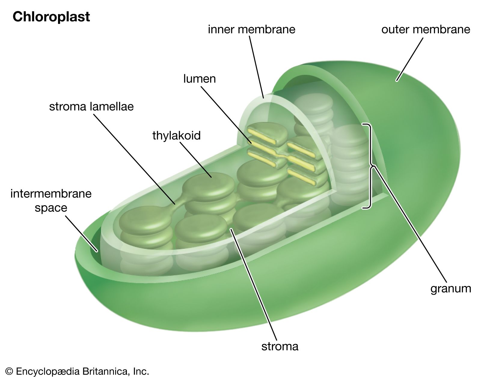
Figure 1: Chloroplast structure [1]
- Thylakoids and Chlorophyll: Among the structures found in chloroplasts are thylakoids – stacks of which are called granum - and embedded in the thylakoid membranes are green pigments called chlorophyll, which are housed in photosystems (functional protein complexes)
- Electron Excitation: When photons hit these chlorophyll molecules, they excite electrons in a photosystem called PSII, and electrons travel along the thylakoid membrane, making it negatively charged and creating an electron transport chain

Figure 2: Chlorophyll absorption spectra [2]
- Proton Movement: Hydrogen ions (H⁺) in the stroma are attracted into the thylakoids as a result of this negatively charged membrane, causing there to be a high concentration of hydrogen in the thylakoids
- Water Splitting (Photolysis): Water molecules in the thylakoids are broken up in a process called photolysis, releasing H⁺, electrons - which replenish the ones that have left PSII - and oxygen which diffuses out of the thylakoid membrane
- Electron Re-excitation: There is another photosystem - PSI - which can capture longer wavelengths of light (680nm) more efficiently than PSII, and PSI re-excites electrons from PSII which have lost energy along the electron transport chain
- NADPH Formation: A cofactor (a sort of 'helper molecule') called NADP⁺ (nicotinamide adenine dinucleotide phosphate) and H⁺ that are both outside of the thylakoid membrane are bonded together by the electrons in the electron transport chain to form NADPH - the reduced state of NADP⁺, helped by an enzyme called ferredoxin-NADP⁺ reductase
- ATP Synthesis: An enzyme called ATP synthase lies across the thylakoid membrane, and H⁺ diffuses from the high concentration inside the thylakoid to the lower concentration in the stroma, and H⁺ helps to bond ADP (adenosine diphosphate) to another phosphate, creating ATP (adenosine triphosphate)
So far, water and sunlight are our inputs, and oxygen, ATP and NADPH are our outputs, and all of these reactions have been light-dependent reactions [3].
Figure 3: Light-dependent reactions of photosynthesis [3]
Now we can move on to the light-independent reactions, known collectively as the Calvin cycle.
- Carbon Fixation: A 5 carbon molecule called ribulose bisphosphate in the stroma bonds with carbon dioxide from the air to create a highly unstable six-carbon intermediate called 2´-carboxy-3-keto-D-arabinitol 1,5-bisphosphate
- Phosphoglycerate Production: The energy contained within ATP and NADPH is used to break this six-carbon intermediate into two molecules of phosphoglycerate, creating ADP and NADP⁺ which are reused for the same purpose
- Regeneration: Some of these phosphoglycerates bond with the help of enzymes to create simple sugars like glucose, and others recombine to make ribulose bisphosphate
- Voila, we have glucose: The glucose created by this process has many uses in the plant, including being stored as starch, providing structural support when linked together as cellulose, and being transported around as sucrose
The Structure of Leaf Tissues
In a biological context, tissues are groups of similar cells that work together to perform a specific function within a plant, and leaves have many different types of tissues.
There are 3 different tissue systems present in the leaf, these are the epidermis, mesophyll tissue (split between the palisade parenchyma and spongy parenchyma), and vascular tissue. Each of these systems contains various types of tissues and cells.
Explore the diagram and descriptions below to learn about the different tissue systems, tissues, cells, structures, and their functions in the leaf:

Figure 4: Leaf tissue structure [4]
Epidermal Tissue
- Cuticle: A waxy cuticle covers the outermost layer of leaves, which slows down water loss from the leaves due to its hydrophobic nature. Cutin and suberin form a matrix, in which long chain fatty acids (waxes) are embedded [5]
- Guard cells: While the cuticle does a stellar job of keeping water out, it also prevents the release of other gases such as oxygen and water vapour. Guard cells line openings called stomata, generally found on the underside of leaves, and control the exchange of gases by forming a stomatal pore. This pore is open when the guard cells are turgid and closed when they are flaccid [6] [7]
- Trichomes: Also known as epidermal hair cells, trichomes are specialised, hair-like outgrowths on the surface of leaves. They perform a variety of functions, such as the protection of leaves from UV radiation, high temperatures and insects, in climates and environments where these pose a threat to the plant [8] [9]
Mesophyll tissue
- Palisade cells: Palisade cells are stacked vertically, are around 1-2 layers thick, and are found in the palisade parenchyma. They are slightly seperated to maximise gas exchange. Palisade cells contain the most chloroplasts of any other type of cell found in the leaves [10]
- Spongy mesophyll cells: Found in the spongy parenchyma directly below the palisade parenchyma, spongy mesophyll cells are irregularly shaped and laid out, though work is being done to further characterise them [11]. Their roles include promoting carbon dioxide absorption from the stomata to the palisade and scattering and absorbing light, but they also contain chloroplasts
Vascular tissue
- Xylem: One of two components that make up the vascular bundle, the role of the xylem is to transport water and some nutrients upwards from the roots to the leaves. [12]. Plants lose a vast amount of water through the stomata, which open to allow carbon dioxide in for photosynthesis, and it is the xylem that replenishes this lost water. Water moves through the leaf stalk (petiole) to the mid-rib, the main thick vein, before branching into smaller veins throughout the leaf
- Phloem: Translocation, the process of moving the products of photosynthesis around the plant, takes place in the phloem, the other component of the vascular bundle. While the xylem is unidirectional, the phloem is bidirectional, transporting sucrose and amino acids in both directions. Sieve tube elements take sucrose from companion cells, and due to a pressure gradient, this sucrose is transported to sink cells in the roots and bulbs, where it is converted into other molecules like starch for storage [13]
References
[1]
[2]
Daniele Pugliesi (Original), M0tty (Vector) (2023). Chlorophyll ab spectra. Wikimedia Commons. View Source
[3]
Samantha Fowler, Rebecca Roush, James Wise (2013). The Light-Dependent Reactions of Photosynthesis. Concepts of Biology, OpenStax. View Source
[4]
[5]
Arya Gulab Chand , Sarkar Sutanni , Manasherova Ekaterina , Aharoni Asaph , Cohen Hagai (2021). The Plant Cuticle: Old Challenges, New Perspectives. Frontiers in Plant Science, 12, 663165. View Source
[6]
Jezek M, Blatt MR. (2017). The Membrane Transport System of the Guard Cell and Its Integration for Stomatal Dynamics. Plant Physiology, 74(2), 487-519. View Source
[7]
Hetherington, A. M., & Woodward, F. I. (2003). The role of stomata in sensing and driving environmental change. Nature, 424, 901-908. View Source
[8]
Ma D, Hu Y, Yang C, et al. (2016). Genetic basis for glandular trichome formation in cotton.. Nat Commun. 2016;7:10456. View Source
[9]
Cao H, Li J, Ye Y, et al. (2020). Integrative Transcriptomic and Metabolic Analyses Provide Insights into the Role of Trichomes in Tea Plant (Camellia Sinensis). Biomolecules. 2020;10(2):311. View Source
[10]
Hejiku A (2025). Structure, Function, and Importance of Palisade Cells in Photosynthesis. J Plant Biochem Physiol. 13:339. View Source
[11]
Borsuk AM, Roddy AB, Théroux-Rancourt G, Brodersen CR. (2022). BStructural organization of the spongy mesophyll. PNew Phytol. 2022;234(3):946-960. View Source
[12]
McElrone, A. J., Choat, B., Gambetta, G. A. & Brodersen, C. R. (2013). Water Uptake and Transport in Vascular Plants. Nature Education Knowledge 4(5):6. View Source
[13]
Connie Rye, Robert Wise, Vladimir Jurukovski, Jean DeSaix, Jung Choi, Yael Avissar (2016). Transport of Water and Solutes in Plants. Biology, OpenStax. View Source